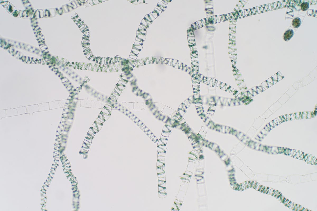
The microscopic world is a fascinating one. As a teacher of biology, it never fails to amaze me when I see a student taking their first view of a living, moving, microscopic organism. As a fishkeeper, I view all manner of tiny beasts that share the aquarium or pond with the fish, plants and inverts that I have. Should the need arise, I can also monitor the populations of those microscopic organisms that live as parasites on and inside the fish.
When using a light microscope, I frequently reflect on how microscopy pioneers must have felt, being the first humans to witness these extraordinary creatures that are usually invisible to the naked eye. Scientists such as Robert Hooke and Anton van Leeuwenhoek lead the way for popularising the viewing of microscopic specimens back in the 1600’s. Books such as Micrographia by Hooke featured detailed drawings of structures invisible to the naked eye for the first time, and Van Leeuwenhoek was the first person to record the presence of tiny moving organisms in a drop of pond water. He used the term ‘animalcules’ to describe the various forms he saw.
Many of these fascinating organisms originate from your pond or aquarium filter, where the waste from your livestock collects and provides a valuable source of food. However, the preparation of a specimen could not be simpler. Turn off the filter of your tank or pond and take foam out of the filter – look for one that is as mucky as possible! Give the foam squeeze in a bucket, add a small amount of tank or pond water and allow the liberated solids to settle. Then using a clean pipette, take a single drop of the mucky water and place onto a microscope slide. Add a cover slip, dab the slide dry with some paper towel and place under the microscope. View at the lowest power first. Even here you will be amazed at the life teeming before your eyes. As you rack up the magnifying power, even more tiny animals will come into view. You will be left with the impression that the fish, plants and inverts really are a tiny fraction of the complex food web of your pond or aquarium.
So, what animals are you likely to see? Microscopic multicellular organisms can easily be seen at 40x magnification, so will often be the first animals to come into focus. Some organisms will already be familiar from the macroscopic world, such as relatives of the earth-worm which share their thick segmented bodies. For me however, viewing organisms is most rewarding when we observe those which have no macroscopic relatives – they belong to some of the less documented branches of the tree of life.
Organisms, such as Rotifers, are fascinating and are often like nothing you’ve seen before! These animals that have paired discs of hair-like cilia at their front end which rhythmically beat giving the appearance of two wheels at the end of the animal. The water current created by these cilia is used by the animal for filter feeding and to aid movement. Like the Rotifers, Gastrotrichs are categorised into their own ‘phylum’ – a broad zoological classification used to categorise species. The Gastrorichs are often called ‘hairy-backs’, and are worm-like with a V-shaped tail and are quite the movers, making them rather tricky to follow around the slide!
Another microscopic animal occasionally seen in a sample from a pond filter is the Tardigrade, or ‘water-bear’ who are also categorised into a phylum of their own. They have eight-legs with small claws at the end of each, hence why they have gained their ‘bear’ comparison. Their black eyes can be clearly seen, and a sword-like stylet is retracted into the head, allowing them to easily feed off of algae cells. Tardigrades are much-loved by biologists, not only for their cute faces and slow lumbering manner, but more so for their ability, when dormant to survive extreme environmental conditions. I frequently take cultures of these organisms into primary schools for children to have their first microscopic encounter.
However, if your microscope has the power, do not stop here! Ramp up the magnification to 100x then 400x to observe more closely the animals described above, but also some much smaller, single-celled organisms will become apparent. Ciliated protozoans such as Paramecium and Euplotes are commonly seen, which have many wavy hairs over their exterior used to keep them moving. They are very mobile and can be tricky to observe closely, but some ciliates are rooted to spot by stalks so are easier to pin down. A bell-shaped body has a ring of beating cilia around the end that creates a current of water so the animal can filter feed. But these organisms aren’t always friendly. Some of these rooted animals can become secondary invaders on fish weakened by a pre-existing disease, giving you as a fishkeeper another good reason to keep an eye on them.
We have only just scratched the surface on what the microorganism world has to offer. I urge all of you to find the time and equipment to observe this life and marvel at its complexity. Like me, you will realise that the fish, plants and inverts you keep in your tanks and ponds are a tiny fraction of this fascinating ecosystem.

